logo Thu, 26 Dec 2024 18:18:18 GMT

Surface Anatomy


Synopsis


British Medical Association Book Awards 2009 - Commended, Basic and Clinical Sciences

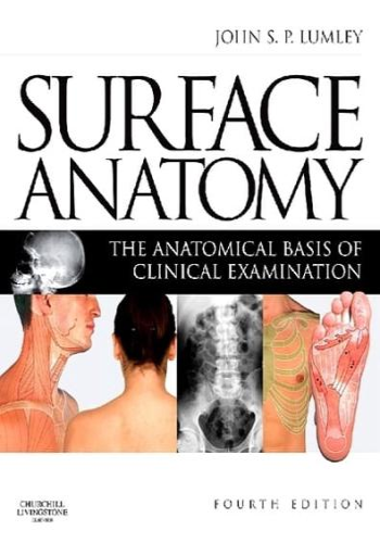
This innovative and highly praised book describes the visible and palpable anatomy that forms the basis of clinical examination. The first chapter considers the anatomical terms needed for precise description of the parts of the body and movements from the anatomical positions. The remaining chapters are regionally organised and colour photographs demonstrate visible anatomy. Many of the photographs are reproduced with numbered overlays, indicating structures that can be seen, felt, moved or listened to. The surface markings of deeper structures are indicated together with common sites for injection of local anaesthetic, accessing blood vessels, biopsying organs and making incisions. The accompanying text describes the anatomical features of the illustrated structures.
  • Over 250 colour photographs with accompanying line drawings to indicate the position of major structures.
  • The seven regionally organised chapters cover all areas of male and female anatomy.
  • The text is closely aligned with the illustrations and highlights the relevance for the clinical examination of a patient.
  • Includes appropriate radiological images to aid understanding.
  • All line drawings now presented in colour to add clarity and improve the visual interpretation.
  • Includes 20 new illustrations of palpable and visible anatomy.
  • Revised text now more closely tied in with the text and with increasing emphasis on clinical examination of the body.

J. S. P. Lumley

Summary

Chapter 1: Introduction to Surface Anatomy

* Summary:
* Importance of surface anatomy in clinical practice
* Different forms of surface anatomy (e.g., palpation, auscultation)
* Relationship between superficial and deep structures
* Example: Palpating the radial pulse (radial artery) on the wrist to assess heart rate and rhythm.

Chapter 2: The Musculoskeletal System

* Summary:
* Muscles, bones, and joints
* Muscle names and functions
* Bone landmarks and their clinical significance
* Joint movements and their associated muscles
* Example: Identifying the deltoid muscle (shoulder abductor) by observing its triangular shape and location on the lateral shoulder.

Chapter 3: The Head and Neck

* Summary:
* Surface anatomy of the face, skull, and neck
* Muscles, bones, and cranial nerves
* Important landmarks for medical procedures (e.g., jugular veins, carotid arteries)
* Example: Locating the mandible (lower jaw) by palpating its inferior border and mandible notch.

Chapter 4: The Thorax and Abdomen

* Summary:
* Muscles, bones, and organs of the thorax and abdomen
* Surface landmarks corresponding to underlying structures
* Respiration, cardiac auscultation, and abdominal examinations
* Example: Auscultating the lungs by listening for breath sounds over the anterior chest wall (auscultatory percussion).

Chapter 5: The Back and Pelvis

* Summary:
* Muscles, bones, and surface landmarks of the back and pelvis
* Spinal cord and peripheral nerves
* Musculoskeletal disorders related to posture and movement
* Example: Palpating the iliac crest (upper edge of the pelvis) to estimate hip bone width.

Chapter 6: The Upper Limb

* Summary:
* Surface anatomy of the arm, forearm, and hand
* Muscles, bones, and nerves
* Joint motions and common injuries
* Example: Locating the median nerve at the wrist by flexing the wrist and observing the muscle contraction in the thenar eminence (base of the thumb).

Chapter 7: The Lower Limb

* Summary:
* Surface anatomy of the hip, thigh, leg, and foot
* Muscles, bones, and joints
* Examination for neurovascular integrity and musculoskeletal injuries
* Example: Testing the Achilles tendon (heel cord) by asking the patient to plantar flex the ankle against resistance.