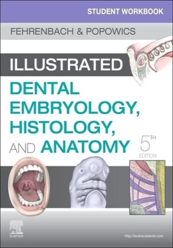
Corresponding to the chapters in Illustrated Dental Embryology, Histology, and Anatomy, 5th Edition, this unique workbook gives you an extensive background in oral biology and the formation and study of dental structures. The fifth edition includes even more case studies with questions presented in the integrated national board format, updated review questions, and removable flashcards to ensure you fully grasp the foundational building blocks of oral healthcare. With updated labeling and terminology exercises, tooth drawing guidelines, and more, this packed resource is an excellent way to prepare for the classroom, board exams, and beyond.
- Comprehensive coverage includes all the content needed for an introduction to the developmental, histological, and anatomical foundations of oral health.
- Detailed case studies include radiographs, clinical photos, profiles, complaints, health histories, and intraoral examination data, each accompanied by multiple-choice questions, to promote critical thinking skills and prepare students for board examinations.
- UNIQUE! Guidelines for Tooth Drawing emphasize fundamental principles in tooth design and include detailed instructions, tips, and dimensions for drawing each permanent tooth.
- Glossary exercises include crossword puzzles and word searches for practice and review of key terminology.
- Expert author Margaret Fehrenbach is one of the most trusted names in dental hygiene education.
- Detachable flashcards help students master tooth morphology and tooth numbering, with multiple-angle drawings of a permanent tooth on one side of the flashcard and characteristics of that tooth on the back.
- A logical organization allows students to focus on areas in which they may need more practice, with units on: (1) anatomic and structure identification and labeling, (2) glossary exercises (3) tooth structure, (4) review questions, and (5) case-based application.
- Perforated workbook pages are three-hole-punched so that they easily fit into a binder, and pages can be removed and submitted to instructors for assignments or extra credit.
- NEW! Expanded structure identification exercises include additional developmental, microbiological, and anatomical structures for more practice identifying and labeling various parts or structures.
- NEW! Review questions for each unit provide even more opportunities for content mastery in preparation for classroom or board exams.
- NEW! Additional case studies include questions using the integrated national board format
- Updated! Evidence-based research thoroughly covers infection control of extracted teeth.
- NEW! Occlusal clinical assessment exercises help prepare students for chairside clinical patient care.


